

Théa Medical Library

Una colección única de libros que abordan diferentes temas y patologías, elaborados en colaboración con oftalmólogos internacionales de reconocido prestigio, líderes en sus respectivas áreas.

Bienvenido a la Théa Medical Library

Su biblioteca médica de referencia en oftalmología, que ofrece una colección digital única creada por expertos internacionales.

La Théa Medical Library es una biblioteca digital cuidadosamente desarrollada por Théa para apoyar la formación y actualización profesional en oftalmología.
Reúne libros y guías clínicas de alta calidad, ricamente ilustrados, redactados en colaboración con reconocidos oftalmólogos de todo el mundo.

Los temas abarcan áreas clave como el OCT y el glaucoma, las úlceras corneales crónicas, los tumores palpebrales y conjuntivales, así como la evolución histórica de los instrumentos y tratamientos oftálmicos.

Disponible a través de Théa Academy, la Biblioteca Médica ofrece a los profesionales del cuidado ocular —desde residentes hasta especialistas experimentados— los recursos necesarios para mantenerse informados sobre los últimos avances clínicos y perspectivas históricas de la especialidad.

Loading…

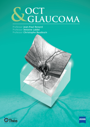
OCT and glaucoma: leveraging imaging for early diagnosis and management

Jean-Paul RENARD / Antoine LABBÉ / Christophe BAUDOUIN,
GlaucomaImagenología oftálmica
Optical Coherence Tomography (OCT) has revolutionized the diagnosis and management of glaucoma, providing clinicians with precise and reproducible imaging of ocular structures. OCT uses light waves to take cross-section pictures of the retina and optic nerve head (ONH). It has become an indispensable tool in the early detection of glaucomatous optic neuropathy, enabling the identification of structural damage before significant visual field defects manifest.
Ver más
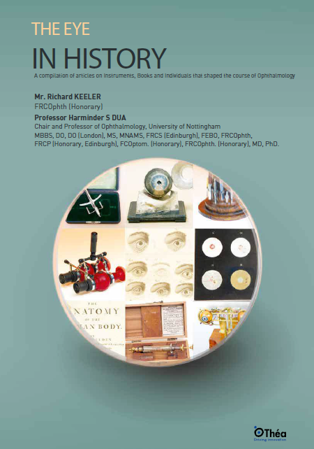
The eye in history: a journey through instruments, diagnostics, and clinical progress

Richard KEELER / Harminder S DUA,
Otros
The evolution of ophthalmology is deeply intertwined with the development of various instruments that have revolutionized the practice. From the early days of the ophthalmoscope, invented by Hermann von Helmholtz in 1850, to the intricate devices used today, each instrument has played a critical role in enhancing our understanding and treatment of the eye. The direct ophthalmoscope, created by Helmholtz, allowed for the first detailed examination of the interior of the eye, laying the groundwork for modern ophthalmology. The subsequent development of the binocular indirect ophthalmoscope by Marc-Antoine Giraud-Teulon and others further expanded the capabilities of eye examination, enabling the visualization of the fundus in three dimensions. These advancements illustrate the shift from theoretical concepts to practical applications in eye history.
Ver más

Practical OCT Angiography: a modern guide to retinal imaging and clinical diagnosis

Sylvia NGHIÊM-BUFFET/ Sandrine AYRAULT / Corinne DELAHAYE-MAZZA / Typhaine GRENET / Gabriel QUENTEL / Franck FAJNKUCHEN / Salomon Yves COHEN,
Imagenología oftálmicaRetina
Optical Coherence Tomography Angiography (OCT-A) is a revolutionary technique in the field of ophthalmology that allows detailed visualization of the retinal and choroidal vascular structures without the need for contrast dye injection. Unlike traditional fluorescein angiography, which involves injecting fluorescein dye into a vein to highlight the blood vessels in your eye, OCT-A uses the movement of red blood cells to create high-resolution scans of the eye’s vascular system. This non-invasive method provides an unprecedented level of detail, allowing clinicians to detect and monitor various eye conditions more effectively.  
Ver más

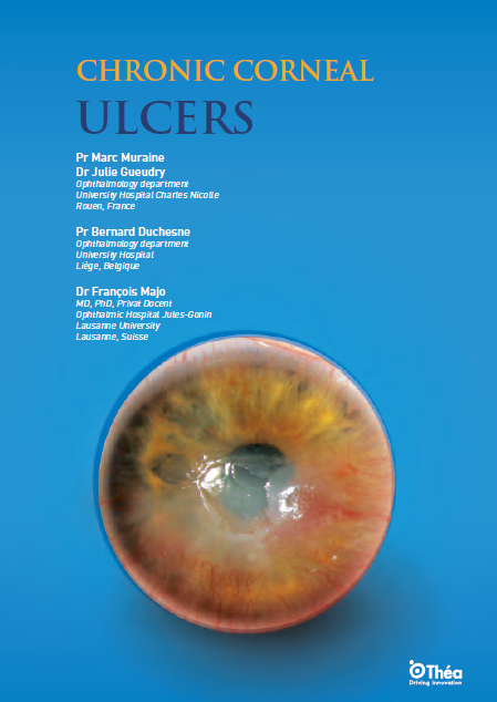
Chronic corneal ulcers: an in-depth review

Marc MURAINE / Julie GUEUDRY / Bernard DUCHESNE / François MAJO,
Córnea
Chronic corneal ulcers are a persistent and debilitating condition characterized by the breakdown of the corneal epithelium with subsequent involvement of the underlying stroma. Unlike acute ulcers, which typically heal within a few days, chronic ulcers do not heal even after three weeks. The pathophysiology of these ulcers is complex and often involves multiple factors that can disrupt the normal healing process of the corneal epithelium.
Ver más

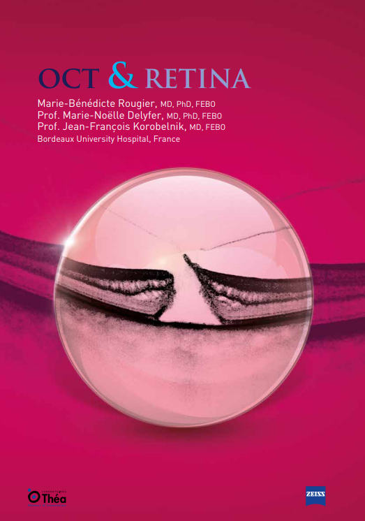
OCT and retina: a comprehensive guide to a macular disease diagnostics

Marie-Bénédicte ROUGIER / Marie-Noëlle DELYFER / Jean-François KOROBELNIK,
Imagenología oftálmicaRetina
Optical Coherence Tomography (OCT) has revolutionized retinal diagnostics over the past 15 years. Through advancements such as Time-Domain OCT (TD-OCT) and Spectral-Domain OCT (SD-OCT), this technology has become accessible to a large number of centers and physicians worldwide. These developments have significantly improved the diagnosis and follow-up of macular diseases, driven by substantial advancements in treatments, especially with the arrival of corticosteroids and anti-VEGF administered by intravitreal (IVT) injections.
Ver más

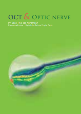
OCT and optic nerve: advancing neuro-ophthalmic imaging

Jean-Philippe NORDMANN,
Imagenología oftálmica
Optical Coherence Tomography (OCT) is a revolutionary imaging technique that has transformed the field of ophthalmology. By using light waves to create detailed cross-sectional images of the retina and optic nerve, OCT has become an indispensable tool in the assessment of optic nerve health and optical nerve imaging. This non-invasive technique allows clinicians to visualize the microstructures within the eye, providing insights that were previously unattainable with conventional imaging methods.
Ver más
Traducir »