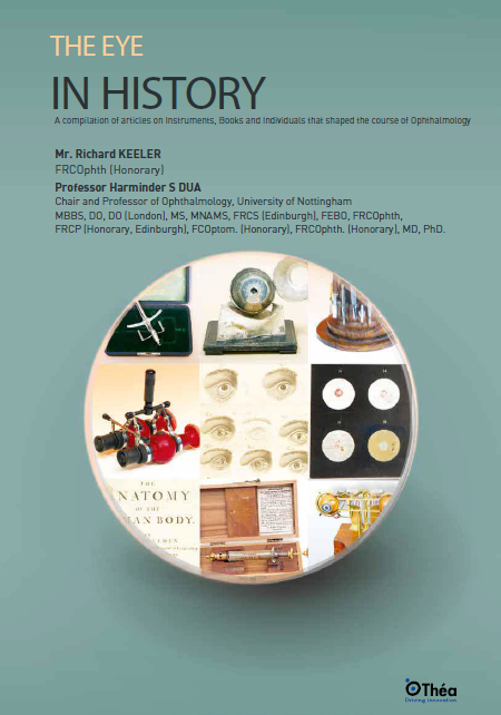
The evolution of ophthalmology is deeply intertwined with the development of various instruments that have revolutionized the practice. From the early days of the ophthalmoscope, invented by Hermann von Helmholtz in 1850, to the intricate devices used today, each instrument has played a critical role in enhancing our understanding and treatment of the eye. The direct ophthalmoscope, created by Helmholtz, allowed for the first detailed examination of the interior of the eye, laying the groundwork for modern ophthalmology. The subsequent development of the binocular indirect ophthalmoscope by Marc-Antoine Giraud-Teulon and others further expanded the capabilities of eye examination, enabling the visualization of the fundus in three dimensions. These advancements illustrate the shift from theoretical concepts to practical applications in eye history.
One significant milestone in the history of ophthalmic instruments was the invention of the slit-lamp biomicroscope by Allvar Gullstrand in the early 20th century. This device enabled detailed examination of the anterior segment of the eye, including the cornea, anterior chamber, and lens. The slit-lamp has become an indispensable tool in the diagnosis and management of various eye conditions, allowing for precise evaluation and documentation of ocular structures. Additionally, the advent of the gonioscope, which allows for the visualization of the anterior chamber angle, further revolutionized the understanding and treatment of glaucoma.
Another noteworthy development was the introduction of the retinoscope, which revolutionized the assessment of refractive errors. The retinoscope allows clinicians to objectively determine a patient’s refractive status by observing the reflection of light from the retina. This instrument, combined with the use of trial lenses and phoropters, has greatly improved the accuracy of vision testing and prescription of corrective lenses. The evolution of these instruments highlights the importance of technological advancements in the field of ophthalmology and their impact on patient care.
The teaching of ocular pathology has seen significant advancements over the centuries. Initially, the use of glass jars to preserve eye specimens was a common practice, providing students with tangible examples of various eye conditions. Maurice Perrin’s introduction of the “phantom eye” in 1866 marked a major advancement in ophthalmic education, allowing students to visualize eye conditions in a more interactive manner. The artificial eye, equipped with various painted conditions, offered a realistic representation of ocular pathology. This method evolved with the advent of photography and digital imaging, which have made it easier to document and study eye diseases. Today, modern teaching aids such as fundus cameras and interactive DVDs have replaced glass jars, ensuring that the memory of ocular pathology is kept alive in the minds of students and practitioners.
The use of virtual reality (VR) and augmented reality (AR) in ophthalmic education has further transformed the learning experience. VR and AR technologies enable students to engage in immersive simulations, providing a realistic and interactive environment for learning. These technologies allow for the visualization of complex ocular structures and pathological conditions, enhancing the understanding of eye anatomy, eye movements and eye diseases. Additionally, the integration of telemedicine and online platforms has facilitated remote learning and collaboration, enabling students and practitioners to access educational resources and consult with experts from around the world.
The establishment of ocular pathology museums and collections has also played a crucial role in preserving the history of eye diseases and treatments. These institutions house extensive collections of eye specimens, photographs, and medical records, providing valuable resources for research and education. Museums such as the Moorfields Eye Hospital Museum in London and the Museum of Vision in San Francisco offer insights into the historical development of ophthalmology and the progress made in the diagnosis and treatment of eye conditions. By preserving and showcasing the history of ocular pathology, these institutions contribute to the ongoing education and advancement of the field.
Vision testing and diagnostics have always been at the heart of ophthalmology. Early instruments like the optometer, designed by William Porterfield in the 18th century, laid the foundation for accurate refractive error measurement. The introduction of cylindrical lenses and the development of optometers with rotating prisms by figures like Emile Javal and Hermann Snellen further refined these diagnostic tools. The invention of the trial lens set by Georg Frönmuller allowed for more precise assessments of visual acuity. Additionally, instruments like the keratometer and ophthalmometer, which measure the curvature of the cornea, have become indispensable in modern ophthalmology. The evolution of these diagnostic tools underscores the importance of accurate vision testing in the field of eye care.
The development of automated perimetry in the mid-20th century marked a significant advancement in the assessment of the visual field. Automated perimetry devices, such as the Humphrey Field Analyzer, enable the systematic and quantitative measurement of peripheral vision. These instruments have become essential in the diagnosis and management of glaucoma, retinal diseases, and neurological conditions affecting the visual pathways. The ability to accurately map the visual field has greatly enhanced our understanding of these conditions and improved patient outcomes.
Advances in imaging technologies have also revolutionized the field of ophthalmic diagnostics. Optical coherence tomography (OCT), introduced in the 1990s, allows for high-resolution, cross-sectional imaging of the retina and optic nerve. OCT has become an indispensable tool in the diagnosis and management of retinal diseases, glaucoma, and other ocular conditions. The development of adaptive optics imaging techniques has further enhanced the resolution and detail of retinal imaging, providing valuable insights into the structure and function of the retina at a cellular level, including color vision. These imaging technologies have greatly expanded our diagnostic capabilities and improved our understanding of ocular diseases.
The history of eye surgery is marked by the contributions of numerous individuals who have advanced the field through their innovations and techniques. Pioneers like Jacques Daviel, who performed the first extracapsular cataract extraction in 1747, set the stage for modern cataract surgery. Figures such as Sir Benjamin Rycroft and Sir Tudor Thomas further refined these techniques, leading to the widespread practice of keratoplasty. Instruments like the von Hippel trephine and the Dimitry instrument revolutionized corneal transplantation and cataract extraction, respectively. The legacy of these individuals and their contributions is evident in the progress made in eye surgery over the centuries. Their dedication to improving surgical outcomes has left an indelible mark on the field, ensuring that the history of eye surgery continues to evolve and improve.
One of the most significant advancements in eye surgery was the development of phacoemulsification by Dr. Charles Kelman in the 1960s. This technique revolutionized cataract surgery by allowing for the removal of the lens through a small incision using ultrasonic energy. Phacoemulsification has become the gold standard for cataract extraction, offering numerous benefits such as faster recovery times, reduced surgical risks, and improved visual outcomes. The introduction of foldable intraocular lenses (IOLs) further enhanced the procedure, allowing
for implantation through the same small incision and providing patients with improved visual acuity.
Advances in laser technology have also had a profound impact on eye surgery. The development of the excimer laser in the 1980s paved the way for laser-assisted in situ keratomileusis (LASIK), a procedure that reshapes the cornea to correct refractive errors. LASIK has become one of the most popular and effective methods for vision correction, offering patients rapid visual improvement and minimal discomfort. The advent of femtosecond lasers has further refined the precision and safety of corneal surgeries, including LASIK and corneal transplants.
The contributions of individuals such as Dr. Patricia Bath, who pioneered the use of laser technology in cataract surgery, and Dr. John Alpar, who developed innovative techniques for corneal transplantation, have further advanced the field of eye surgery. Their dedication to improving surgical outcomes and patient care has left a lasting legacy, inspiring future generations of ophthalmologists to continue pushing the boundaries of what is possible in eye surgery.
In conclusion, the history of ophthalmology is a rich tapestry of innovation, education, and surgical advancements. The development of instruments, the evolution of teaching methods, the foundation of vision testing, and the contributions of pioneering surgeons have collectively shaped the field. By understanding the past, we can better appreciate the progress made and continue to build on the legacy of those who have dedicated their lives to the study and treatment of the eye.


