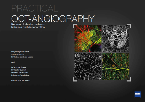
Optical Coherence Tomography Angiography (OCT-A) is a revolutionary technique in the field of ophthalmology that allows detailed visualization of the retinal and choroidal vascular structures without the need for contrast dye injection. Unlike traditional fluorescein angiography, which involves injecting fluorescein dye into a vein to highlight the blood vessels in your eye, OCT-A uses the movement of red blood cells to create high-resolution scans of the eye’s vascular system. This non-invasive method provides an unprecedented level of detail, allowing clinicians to detect and monitor various eye conditions more effectively.
OCT-A operates using light waves to take cross-sectional images of the retina, which are then compiled to form a detailed map of the blood flow within the eye. This technique allows for the observation of microvascular changes that are often invisible with conventional imaging methods. The ability to visualize retinal and choroidal blood vessels in such detail has greatly impacted the management of diseases such as glaucoma, diabetic retinopathy, and age-related macular degeneration (AMD). With OCT-A, clinicians can identify abnormal blood flow, monitor disease progression, and evaluate the effectiveness of treatments with remarkable precision.
Furthermore, OCT-A has become an essential tool in research, advancing our understanding of various ocular diseases and conditions. Researchers have utilized OCT-A to study the early signs of vascular changes in the retina, providing insights that could lead to the development of new therapeutic strategies. The detailed imaging capability of OCT-A has also facilitated the exploration of genetic factors influencing ocular health, contributing to a broader understanding of the pathophysiology of eye diseases.
Fluorescein angiography has long been a staple in eye treatment, utilizing a special camera to capture the flow of fluorescein dye as it moves through the blood vessels in the eye. While effective, this method has several limitations, including potential side effects such as allergic reactions and sensitivity to light, which occur when the dye leaks during the injection. In contrast, OCT-A offers a safer alternative by eliminating the need for dye injection, thereby reducing the risk of allergic reactions and other side effects. OCT-A’s high-resolution scans provide a more detailed view of the retinal and choroidal vessels, making it an invaluable tool in diagnosing and monitoring conditions such as diabetic retinopathy, macular degeneration, and retinal vein occlusion.
The comparison between OCT-A and fluorescein angiography highlights the advancements that OCT-A has brought to the field. Fluorescein angiography involves that fluorescein is injected in a vein, which travels through the bloodstream to the blood vessels in the eye. A special camera then captures images of the dye as it moves through these vessels, allowing clinicians to identify areas of leakage, blockage, or abnormal growth. While fluorescein angiography provides valuable information, it comes with risks such as allergic reaction to the dye, sensitivity to light, and other side effects. Patients may experience nausea, vomiting, or in rare cases, anaphylactic shock.
OCT-A, on the other hand, does not require any dye injections, making it a much safer option for patients. The technique relies on the movement of red blood cells to map the blood flow within the eye, providing detailed images without the need for invasive procedures. This non-invasive nature of OCT-A makes it particularly advantageous for patients with allergies or those who are sensitive to the dye used in fluorescein angiography. Additionally, OCT-A’s high-resolution scans offer a more comprehensive view of the retinal and choroidal vessels, enhancing the ability to detect and monitor ocular conditions.
In clinical settings, the adoption of OCT-A has led to improved patient outcomes and streamlined diagnostic processes. For instance, in diabetic retinopathy, OCT-A allows for the detailed visualization of microaneurysms, non-perfusion areas, and neovascularization without the need for fluorescein dye. This capability is particularly useful in detecting early stages of the disease and monitoring treatment efficacy. Similarly, in age-related macular degeneration (AMD), OCT-A provides clear images of the abnormal blood vessels growing beneath the retina, which are crucial for determining the severity and progression of the condition. The technique’s ability to produce high-resolution images without the risks associated with dye injections makes it a preferred choice for both patients and clinicians.
In clinical practice, OCT-A has shown significant advantages over traditional fluorescein angiography. For instance, in diabetic retinopathy, OCT-A allows for the detailed visualization of microaneurysms, non-perfusion areas, and neovascularization without the need for fluorescein dye. This capability is particularly useful in detecting early stages of the disease and monitoring treatment efficacy. Similarly, in age-related macular degeneration (AMD), OCT-A provides clear images of the abnormal blood vessels growing beneath the retina, which are crucial for determining the severity and progression of the condition. The technique’s ability to produce high-resolution images without the risks associated with dye injections makes it a preferred choice for both patients and clinicians.
The practical applications of OCT-A are extensive, ranging from diagnosis to ongoing monitoring of various ocular conditions. For example, in glaucoma management, OCT-A can be used to assess changes in the optic nerve head and surrounding vascular structures. By providing detailed images of blood flow within the eye, OCT-A enables clinicians to detect early signs of glaucomatous damage and monitor disease progression over time. This information is critical for adjusting treatment plans and preventing further vision loss.
In addition to its diagnostic capabilities, OCT-A has proven to be invaluable in surgical planning and post-operative care. For patients undergoing retinal surgery, OCT-A can be used to map the retinal vasculature, guiding surgeons in their approach and ensuring precise interventions. Post-surgery, OCT-A facilitates the monitoring of healing and the detection of potential complications such as retinal detachment or neovascular growth. The ability to visualize blood flow in such detail aids in the timely and effective management of surgical outcomes.
Furthermore, OCT-A has found applications in pediatric ophthalmology, offering a non-invasive method to examine vascular changes in children with inherited or developmental ocular conditions. The technique’s ability to provide high-resolution images without the need for dye injections is particularly beneficial for young patients who may be sensitive or uncooperative during invasive procedures. OCT-A’s detailed imaging supports early diagnosis and intervention, improving long-term outcomes for pediatric patients.
The advent of OCT-A marks a significant milestone in the field of ophthalmology, offering clinicians a powerful tool for diagnosing and monitoring a wide range of retinal and choroidal conditions. Its non-invasive nature, combined with the ability to produce high-resolution scans, significantly enhances the accuracy and safety of eye examinations.


