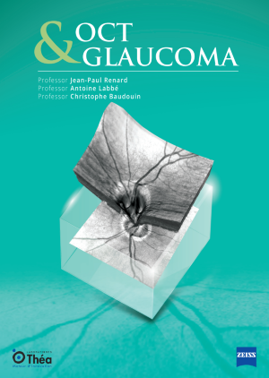
Optical Coherence Tomography (OCT) has revolutionized the diagnosis and management of glaucoma, providing clinicians with precise and reproducible imaging of ocular structures. OCT uses light waves to take cross-section pictures of the retina and optic nerve head (ONH). It has become an indispensable tool in the early detection of glaucomatous optic neuropathy, enabling the identification of structural damage before significant visual field defects manifest.
The advent of OCT technology has allowed eye care professionals to visualize the intricate layers of the retina and the optic nerve head with unprecedented detail. This non-invasive imaging technique helps in detecting early signs of glaucoma, even before patients experience noticeable vision loss. By analyzing the structural changes in these ocular components, clinicians can diagnose glaucoma at a much earlier stage compared to traditional methods. Early detection is crucial in glaucoma management as it allows for timely intervention and treatment, which can slow down or prevent further progression of the disease.
Moreover, OCT provides quantitative data that is highly valuable for monitoring changes over time. The ability to measure the thickness of the RNFL (Retinal Nerve Fibre Layer) and other retinal layers accurately enables clinicians to track the progression of glaucoma in a precise manner. This is particularly important for tailoring individualized treatment plans and making informed decisions about the need for therapeutic adjustments. In summary, the role of OCT in glaucoma detection cannot be overstated, as it offers a powerful means to identify and manage this potentially blinding condition effectively.
One of the primary indicators of glaucoma is the thinning of the Retinal Nerve Fibre Layer (RNFL). OCT allows for the detailed examination of the RNFL, which is crucial for assessing the extent of glaucomatous damage. By providing high-resolution images, clinicians can observe changes in the optic nerve head (ONH) and detect characteristic glaucomatous alterations such as rim thinning and increased cupping. The ability of OCT to measure these changes quantitatively aids in distinguishing glaucoma from other optic neuropathies.
The RNFL is composed of the axons of ganglion cells, which are the nerve cells responsible for transmitting visual information from the retina to the brain. In glaucoma, these axons are damaged, leading to RNFL thinning, which can be detected using OCT. The early detection of RNFL thinning is essential in glaucoma management as it allows for the implementation of treatment strategies before significant vision loss occurs.
Additionally, OCT can detect changes in the optic nerve head, such as increased cupping and rim thinning, which are characteristic features of glaucomatous damage. These structural changes can be monitored over time to assess the progression of the disease. By providing a detailed view of the RNFL and ONH, OCT enables clinicians to make accurate diagnoses and track the effectiveness of treatment strategies.
Visual field defects are a hallmark of glaucoma, but they usually appear only after significant RNFL damage has occurred. OCT enables the correlation of structural changes with functional impairment by comparing RNFL thickness and ONH parameters with visual field test results. This correlation is vital for confirming glaucoma diagnosis and understanding the progression of the disease. Identifying areas of RNFL thinning on OCT that correspond to visual field defects can help in tailoring individualized management plans for patients.
The relationship between structural changes and functional impairment in glaucoma is complex. While visual field tests assess the functional aspect of vision, OCT provides a structural analysis of the retina and optic nerve head. By correlating these two types of data, clinicians can gain a comprehensive understanding of the disease and its progression. This holistic approach allows for more accurate diagnoses and better-informed treatment decisions.
Furthermore, OCT can help identify pre-perimetric glaucoma, a stage of the disease where structural damage is present, but visual field defects have not yet appeared. Detecting glaucoma at this early stage is crucial for preventing further progression and vision loss. By combining OCT findings with visual field tests, clinicians can develop a more complete picture of the patient’s condition and provide more effective care.
OCT is invaluable for monitoring the progression of glaucoma over time. By performing serial OCT scans, clinicians can detect subtle changes in RNFL thickness and ONH parameters, which indicate disease progression. This longitudinal analysis is essential for adjusting treatment strategies to prevent further vision loss. Advanced OCT devices now offer progression analysis software that compares current scans with baseline measurements, highlighting significant changes and aiding in timely intervention.
The ability to monitor glaucoma progression with OCT is particularly important in managing chronic cases of the disease. As glaucoma is a lifelong condition, ongoing monitoring is essential to ensure that treatment strategies remain effective. Serial OCT scans provide a detailed record of changes in the RNFL and ONH, allowing clinicians to track the disease’s progression accurately.
By identifying subtle changes in the RNFL and ONH, clinicians can adjust treatment plans as needed to prevent further damage and vision loss. This proactive approach to glaucoma management is essential for maintaining the patient’s quality of life and preserving their vision. The use of advanced OCT devices and progression analysis software further enhances the ability to monitor glaucoma progression accurately and make timely interventions.
In conclusion, Optical Coherence Tomography has transformed glaucoma care by providing detailed insights into the structural changes associated with the disease. The ability to detect RNFL thinning, correlate findings with visual field defects, and monitor progression ensures that clinicians can offer the best possible care to their patients. As OCT technology continues to evolve, its role in glaucoma diagnosis and management will only become more significant, ultimately enhancing patient outcomes and preserving vision.
The advancements in OCT technology have led to more precise and reliable imaging, making it an indispensable tool in glaucoma management. By providing detailed and quantitative data on the RNFL and ONH, OCT allows for early detection, accurate diagnosis, and effective monitoring of glaucoma progression. These insights are crucial for developing individualized treatment plans and ensuring that patients receive the best possible care.
As the understanding of glaucoma continues to grow, the use of OCT will remain at the forefront of diagnostic and monitoring techniques. Its ability to provide detailed structural information, combined with its non-invasive nature, makes it an ideal tool for managing this complex and potentially blinding condition. By enhancing glaucoma care with OCT insights, clinicians can help preserve vision and improve the quality of life for their patients.


