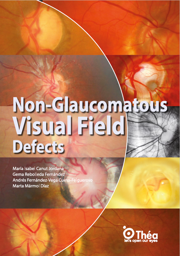
Non-glaucomatous visual field defects are alterations in visual perception not caused by glaucomatous damage to the optic nerve. This book highlights the importance of distinguishing these from typical glaucomatous field loss, as they often stem from neuro-ophthalmological or congenital conditions, each requiring distinct diagnostic and therapeutic strategies. Common conditions producing these defects include optic disc drusen, tilted disc syndrome, optic nerve hypoplasia, optic disc coloboma, and morning glory disc anomaly.
These conditions may be associated with systemic syndromes, developmental anomalies, or genetic disorders, and often present with unique visual field patterns. Misidentifying them as glaucoma can lead to inappropriate treatments and missed systemic diagnoses. The book uses OCT, visual field testing, and neuroimaging to clarify these entities, stressing careful analysis and differential diagnosis.
The book details several congenital and acquired neuro-ophthalmological conditions that can mimic glaucomatous damage:
- Optic Disc Drusen: Calcified deposits in the optic nerve head, often bilateral and asymptomatic. They cause visual field defects, especially arciform or enlarged blind spots, and may mimic papilledema. EDI-OCT and autofluorescence are key diagnostic tools.
- Tilted Disc Syndrome (TDS): A congenital disc anomaly often associated with high myopia and astigmatism. The disc appears obliquely oriented, frequently causing pseudo-hemianopia and other defects. OCT helps assess peripapillary structures and avoids misdiagnosis with glaucoma.
- Optic Nerve Hypoplasia (ONH): A leading cause of childhood blindness. Presents with a small, pale optic disc and the characteristic “double ring sign.” Associated with developmental, endocrine, and neuroanatomical anomalies. Visual fields vary from normal to severe loss.
- Optic Disc Coloboma: A rare congenital excavation of the optic disc due to incomplete embryonic fissure closure. May present alone or with other ocular/systemic colobomas. Visual loss depends on papillomacular involvement. OCT and fluorescein angiography assist in differentiation.
- Morning Glory Disc Anomaly: A large, excavated optic disc with a central glial tuft and radiating vessels. Often associated with retinal detachment and severe field loss. It may signal cerebrovascular or syndromic conditions like Moyamoya or PHACE syndrome.
Each pathology is illustrated with imaging and case studies, supporting precise differential diagnosis.
Accurate diagnosis of non-glaucomatous visual field defects relies on combining visual field testing, fundus examination, and advanced imaging. OCT (Optical Coherence Tomography) plays a central role, particularly EDI OCT and swept-source OCT, which enhance visualization of deep optic nerve structures and distinguish buried optic disc drusen from papilledema.
Visual field tests, such as Humphrey perimetry, reveal characteristic defects enlarged blind spots, altitudinal or hemianopic patterns that differ from glaucomatous cupping. However, interpretation requires caution to avoid mislabeling congenital anomalies as progressive glaucomatous loss.
Neuroimaging, including MRI and CT, is crucial for systemic conditions like optic nerve hypoplasia or morning glory disc, particularly when endocrinopathy or brain malformations are suspected. OCT-Angiography (OCTA) and PHOMS (peripapillary hyperreflective ovoid mass-like structures) detection are newer tools that aid in distinguishing neuro-ophthalmic from glaucomatous causes.
OCT also allows for minimum rim width (MRW) and ganglion cell complex analysis metrics that provide better accuracy in tilted discs or high myopia, where RNFL data may be unreliable.
This book is an essential tool for EBO exam preparation, offering a case-based, image-rich review of non-glaucomatous visual field defects. It presents high-yield differential diagnosis frameworks, distinguishing structural anomalies from glaucomatous patterns. Each condition includes clinical, imaging, and visual field correlation, strengthening diagnostic reasoning.
For young ophthalmologists, especially those preparing for the European Board of Ophthalmology (EBO) exam, this resource builds confidence in managing rare visual field defects and reduces diagnostic uncertainty. It complements other neuro-ophthalmology texts by translating theory into practice with real clinical examples and annotated OCTs.
Understanding non-glaucomatous visual field defects is crucial for accurate diagnosis and management of optic nerve pathologies. This book provides a comprehensive, clinically grounded overview of congenital and acquired anomalies that can mimic glaucoma. With case illustrations, OCT findings, and diagnostic algorithms, it equips clinicians with the tools to improve patient outcomes.
To explore the full content, ophthalmologists are encouraged to log in or register on Théa Academy. The Théa Medical Library offers access to this and other leading resources in neuro-ophthalmology and glaucoma management, keeping professionals updated with current diagnostic and therapeutic strategies.


