Ocular and orbital ultrasound is a non-invasive imaging technique that utilizes high frequency sound waves to visualize the structures of the eye and surrounding orbital tissues. This method is particularly valuable when direct examination is hindered by media opacities, such as dense cataracts, vitreous hemorrhages, or corneal scars.
With ocular and orbital ultrasound, clinicians can examine every part of the eye, from the cornea through to the optic disc, as well as assess the orbit itself. This comprehensive visualization is crucial for detecting and evaluating eye injuries, locating foreign bodies, and monitoring conditions that may indicate increased intracranial pressure (such as optic nerve sheath diameter changes).
Additionally, this imaging technique is indispensable in the pre- and post-operative evaluation of patients, especially in cataract surgery. It helps in the assessment of the posterior segment when the view is obstructed and is essential for verifying the correct positioning of a lens implant after surgery.
In summary, ocular and orbital ultrasound offers a safe, efficient, and detailed approach to diagnosing and managing a wide range of ocular and orbital conditions, making it a cornerstone in modern ophthalmic practice.
Ocular ultrasonography is a cornerstone diagnostic tool in modern ophthalmology, offering rapid, non-invasive assessment for a wide range of conditions. It is particularly valuable for diagnosing conditions including retinal detachment, posterior vitreous detachment, choroidal tumors, and various optic nerve anomalies. Thanks to its ability to visualize fluid-filled spaces within the eye, ocular ultrasonography is highly effective even when direct visualization is obstructed by media opacities.
Both B-mode and standardized A-mode techniques are used in eye and orbit ultrasound. B-mode provides detailed cross-sectional images, allowing for dynamic assessment of ocular structures and the detection of subtle changes in real time. A-mode is especially useful for precise measurements, such as axial length, which is critical for intraocular lens (IOL) calculations.
Ocular ultrasonography also plays a key role in evaluating intraocular lens positioning after cataract surgery, guiding treatment decisions in cases of ocular trauma, and monitoring the progression of diseases affecting the posterior segment. Recent advances, such as the introduction of annular probe technologies and 20 MHz systems, now enable ultra-high resolution imaging of both the anterior and posterior segments of the eye.
In summary, ocular ultrasonography is indispensable for diagnosing, managing, and tracking a variety of ocular and orbital conditions, ensuring accurate, real-time insights that directly impact patient care.
Eye and orbit ultrasound is an indispensable tool throughout the continuum of ophthalmic care, from initial diagnosis to surgical planning and follow-up. In situations where optical coherence tomography (OCT) or slit-lamp examination is not possible—such as with dense cataracts, vitreous hemorrhage, or corneal opacities—ocular and orbital ultrasound becomes the primary imaging modality.
This technology is especially valuable in cataract surgery planning. It allows clinicians to assess the posterior segment of the eye when direct visualization is obscured and to accurately measure axial length, which is essential for selecting and positioning the appropriate lens implant. Ultrasound also plays a key role in the early detection of changes in the optic nerve and retina, enabling timely intervention for conditions such as retinal detachment or optic nerve anomalies.
The book highlights the advantages of standardized echography, which enhances diagnostic precision by helping to differentiate between various intraocular tumors, measure ocular structures, and confirm complex diagnoses. This level of detail is crucial for tailoring treatment plans, minimizing surgical risks, and optimizing patient outcomes.
In summary, eye and orbit ultrasound bridges the gap between clinical examination and advanced imaging, supporting every stage of eye care, from diagnosis and surgical planning to post-operative assessment, ensuring accurate, safe, and effective management of ocular conditions.
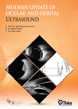
This modern update on ocular and orbital ultrasound is an essential educational resource for both clinicians and students. The book stands out by combining clear, step-by-step imaging methodology with a comprehensive atlas of real-life ultrasound cases, making complex concepts accessible to practitioners at every level.
It offers detailed technical guidance on performing and interpreting ocular ultrasonography, with practical examples that span a wide range of clinical scenarios. Readers will find in-depth protocols for using eye and orbit ultrasound in the diagnosis and management of retinal detachment, evaluation of optic disc abnormalities, and the assessment of intraocular lens positioning or complications. Each case is illustrated with high-quality images, helping users build confidence in recognizing key ultrasound findings.
Designed to foster best practices, the book encourages the routine integration of ocular ultrasound into ophthalmic care, emphasizing its value in both emergency and routine settings. It also serves as an ideal companion to Théa Academy’s broader ocular imaging training resources, supporting continuous learning and professional development.
In summary, this book is a practical and authoritative guide for anyone seeking to master ocular and orbital ultrasound, making it an invaluable addition to the educational toolkit of ophthalmologists, residents, and medical students alike.