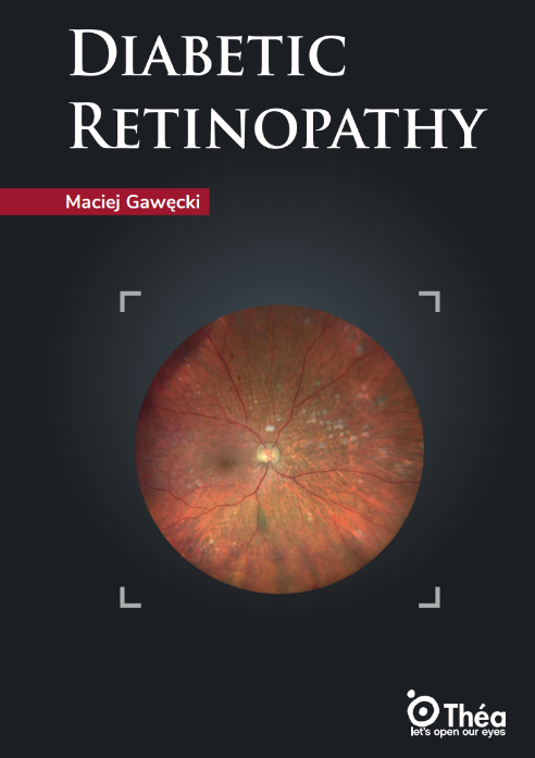
Diabetic retinopathy is one of the most serious complications of diabetes and a leading diabetes-related eye disease, affecting the retina, the delicate tissue at the back of the eye that is essential for vision. High blood sugar levels over time cause progressive retinal damage by harming the small blood vessels in the retina, leading them to leak fluid or bleed. This process can result in swelling (macular edema), the formation of abnormal new blood vessels (proliferative diabetic retinopathy), and, eventually, progressive vision impairment.
The risk and severity of diabetic retinopathy are closely linked to the age at which diabetes is diagnosed and the duration of the disease. Studies have shown that diabetic retinopathy, as well as its more severe forms, proliferative diabetic retinopathy and diabetic macular edema, are more frequent in patients diagnosed with diabetes before the age of 30. This is particularly true for individuals who require insulin therapy, as they often have type 1 diabetes and live with the disease for many years. In contrast, those diagnosed later in life, especially those not needing insulin, tend to have a lower risk of developing these vision-threatening complications.
Early stages of this diabetes-related eye disease often have no symptoms, making regular eye examinations crucial for all people with diabetes. As the disease progresses, patients may notice blurred vision, dark spots, or even sudden vision loss. Without timely intervention, diabetic retinopathy can lead to irreversible blindness.
Early diagnosis and management are essential to prevent severe vision loss caused by retinal damage. Regular retinal screenings, strict blood sugar control, and prompt treatment, such as laser therapy or injections, can slow the progression of the disease and preserve vision. Therefore, proactive management and close monitoring are key to reducing the impact of diabetic retinopathy on vision and maintaining quality of life for people with diabetes.
Diabetic retinopathy arises as a result of complex interactions between metabolic, genetic, and environmental factors in individuals with diabetes. Prolonged periods of elevated blood sugar, known as hyperglycemia, play a central role in damaging the tiny blood vessels that supply the retina. Over time, this damage compromises the retina’s ability to function properly, making diabetic retinopathy one of the most common and serious diabetic eye complications. Hypertension, or high blood pressure, further increases the risk by putting additional strain on these delicate vessels, accelerating the progression of retinal injury and increasing the likelihood of vision loss.
Genetic predisposition also influences an individual’s susceptibility to diabetic retinopathy, with some people being more vulnerable to retinal changes than others. Beyond these main causes, several risk factors can speed up the development and severity of the disease. These include the type of diabetes, insulin dependence, duration of diabetes, and the age at which diabetes is diagnosed. Lifestyle factors such as poor diet, lack of physical activity, and limited access to healthcare or education can also contribute to earlier onset and more rapid progression of diabetic eye complications. Additionally, certain life stages, like puberty and pregnancy, are known to increase the risk of developing retinopathy. Together, these factors highlight the importance of comprehensive diabetes management and regular eye examinations to prevent vision loss and protect eye health.
Diabetic retinopathy (DR) is a progressive eye disease classified into stages, starting with nonproliferative diabetic retinopathy (NPDR) and advancing to proliferative diabetic retinopathy (PDR).
NPDR is the early stage, characterized by microaneurysms, retinal hemorrhages, and vascular changes without new blood vessel growth. As NPDR progresses from mild to moderate and severe forms, retinal blood vessels become increasingly damaged, leading to leakage and retinal swelling. Vision may remain unaffected in early NPDR, but as the disease advances, patients can experience blurred vision and an increased risk of vision-threatening complications.
PDR represents the most advanced stage, marked by the proliferation of new, fragile blood vessels on the retina and into the vitreous. These abnormal vessels can bleed, causing vitreous hemorrhage, retinal detachment, and significant vision loss or even blindness. Disease progression from NPDR to PDR is influenced by factors such as diabetes duration, glycemic control, and hypertension.
Epidemiological studies estimate that about 35.4% of diabetic patients develop some form of retinopathy (NPDR or PDR), with PDR affecting approximately 7.5%. The prevalence is higher in type 1 diabetes (NPDR/PDR: 77.3%/32.4%) compared to type 2 diabetes (25.2%/3%), underscoring the importance of early detection and management to prevent vision loss.
Detecting diabetic retinopathy, a major diabetes-related eye disease, relies on several complementary diagnostic tools to identify and monitor retinal changes. The foundation is the fundus examination, performed by an ophthalmologist using a slit lamp and specialized lenses. This allows direct visualization of the retina to detect microaneurysms, hemorrhages, exudates, and neovascularization, hallmarks of diabetic retinopathy, even before symptoms appear.
Fluorescein angiography is a key imaging technique where a fluorescent dye is injected into the bloodstream, highlighting retinal blood vessels on sequential photographs. This test reveals areas of vascular leakage, ischemia, and neovascularization, providing crucial information on disease severity and guiding treatment decisions.
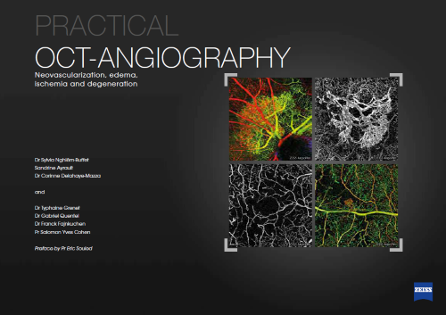
The OCT scan (optical coherence tomography) is now indispensable in diabetic retinopathy care. Modern OCT devices, including spectral-domain (SD-OCT) and swept-source (SS-OCT), generate high-resolution, cross-sectional images of the retina at remarkable speed. OCT can precisely detect and quantify diabetic macular edema, visualize subtle structural changes, and monitor response to therapy. OCT angiography (OCTA), a newer advancement, can non-invasively map retinal blood flow and highlight ischemic zones without dye injection.
Together, these diagnostic tools enable early detection, accurate staging, and close monitoring of diabetic retinopathy, helping to preserve vision in people with diabetes.
Treatment for diabetic retinopathy involves several complementary approaches, tailored to the stage and severity of the disease. Laser therapy is a standard treatment, particularly for proliferative diabetic retinopathy and diabetic macular edema. The laser can destroy ischemic retinal areas or seal leaking vessels, thereby limiting the progression of neovascularization and reducing the risk of hemorrhage or retinal detachment.
Intravitreal injections of anti-VEGF agents or corticosteroids have become essential, especially for diabetic macular edema and early proliferative forms. The objectives of those treatments are to reduce edema, inhibit the growth of abnormal vessels, and improve visual acuity, though they often require repeated injections.
Vitrectomy is reserved for advanced stages, such as persistent vitreous hemorrhage, tractional retinal detachment, or failure of other treatments. This surgical procedure involves removing the vitreous and directly treating retinal complications, allowing for vision restoration or preservation in severe cases. The selection and combination of these treatments depend on the evolutionary stage of diabetic retinopathy and the observed lesions.
Reducing the risk of vision loss from diabetic retinopathy requires a proactive and comprehensive approach. The most effective prevention of diabetic retinopathy begins with strict control of blood sugar levels, as sustained hyperglycemia is the main driver of retinal damage. Maintaining optimal blood pressure and lipid profiles is equally crucial, as hypertension and dyslipidemia accelerate the progression of diabetes-related eye disease.
Regular ophthalmologic examinations are essential, even in the absence of symptoms, since early stages of diabetic retinopathy are often asymptomatic. Annual fundus exams allow for early detection and timely intervention, significantly improving the chances of preserving vision.
Lifestyle modifications play a central role in prevention. Adopting a balanced diet, engaging in regular physical activity, and avoiding smoking all contribute to better metabolic control and overall vascular health. Managing stress, ensuring adequate sleep, and addressing comorbidities such as obesity or vitamin deficiencies further reduce the risk of complications.
In summary, the key to the prevention of diabetic retinopathy and vision loss lies in rigorous metabolic control, healthy living, and regular eye screening. For more detailed insights and practical resources on diabetic eye disease, discover further materials on the Théa Academy website.


