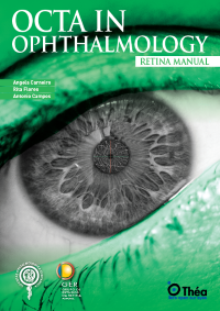
Optical Coherence Tomography Angiography (OCTA) is a cutting-edge, non-invasive method that enables detailed visualization of the retinal and choroidal microvasculature without the need for dye injections. OCTA in ophthalmology has emerged as a revolutionary imaging technique, providing depth-resolved, high-resolution imaging of retinal blood flow by detecting motion contrast from red blood cells. Unlike traditional fluorescein angiography (FA), which is limited to
two-dimensional imaging and is affected by leakage and staining, optical coherence tomography angiography allows clinicians to assess both the structure and function of the eye’s microvascular networks in real time.
The technology operates by acquiring repeated B-scans at the same location and analyzing differences to detect blood flow. Various algorithms, such as split-spectrum amplitude decorrelation angiography (SSADA) and optical microangiography (OMAG), are employed to generate OCTA images. These innovations allow segmentation of individual vascular layers, including the superficial, deep, and choroidal plexuses. OCTA thus enables the visualization of previously inaccessible vascular structures, aiding in early diagnosis and monitoring of ocular diseases.
OCTA is superior in many aspects to traditional methods, especially in avoiding artifacts caused by dye leakage and providing clearer images in cases of hemorrhage or media opacities. However, the technology does have limitations such as motion artifacts and limited field of view, which are being addressed through improvements in image acquisition and processing.
OCTA in ophthalmology has become a valuable tool in the diagnosis and management of various retinal vascular diseases. It plays a crucial role in identifying and monitoring pathologies such as diabetic retinopathy, age-related macular degeneration (AMD), and retinal vein and artery occlusions. The technology enables the detection of microvascular abnormalities like capillary non-perfusion, microaneurysms, and areas of ischemia with high precision.
In diabetic retinopathy imaging, OCTA is particularly useful in visualizing the foveal avascular zone and evaluating changes in vascular density. It helps distinguish between non-proliferative and proliferative stages, and in monitoring response to anti-VEGF therapies. For choroidal neovascularization seen in AMD, OCTA provides a clear view of neovascular membranes, allowing subtype classification and assessment of treatment efficacy over time.
Additionally, OCTA aids in detecting early signs of retinal vascular occlusions by identifying ischemic areas and structural changes that are not always visible with conventional imaging. The ability to perform repeated scans without contrast agents supports ongoing patient monitoring and enhances disease management.
By offering a detailed, layer-by-layer view of retinal and choroidal circulation, OCTA facilitates a better understanding of disease mechanisms and supports more targeted therapeutic interventions.
OCTA interpretation requires careful attention to anatomical detail and awareness of common pitfalls. Clinicians must differentiate between normal vascular structures and pathological patterns. Key features such as the foveal avascular zone, vessel density, and capillary dropout should be assessed. OCTA also supports retinal blood flow analysis through en face imaging and cross-sectional views with flow overlays.
A practical approach includes recognizing artifacts such as projection artifacts, motion distortions, and segmentation errors, which can mislead interpretation. Motion correction technologies and enhanced algorithms are increasingly used to improve image reliability.
Clinicians should be cautious in evaluating images, especially in eyes with structural anomalies or media opacities. Adjusting segmentation boundaries and comparing multiple scan sizes help optimize interpretation. Overall, OCTA enhances clinical decision-making by offering objective data and high-resolution images of microvascular health.
Despite its benefits as a non-invasive method, OCTA in ophthalmology has certain technical limitations. These include a restricted field of view, sensitivity to motion artifacts, and challenges in segmentation accuracy, particularly in patients with retinal distortion from disease. Some vascular changes, like leakage, are not visible with OCTA, unlike with FA (Fluorescein Angiography ) or ICGA (Indocyanine Green Angiography).
Another limitation is the two-dimensional representation of complex three-dimensional vascular networks, which can hinder accurate interpretation. Moreover, variability among devices and algorithms can affect reproducibility and comparability of results.
Looking forward, ongoing research aims to overcome these challenges by developing wide-field imaging, 3D visualization tools, and deep learning-based artifact correction algorithms. Enhanced speed and resolution in newer devices will also contribute to broader clinical applicability and more accurate diagnosis of retinal diseases.
Integrating OCTA in ophthalmology into routine clinical workflows enhances diagnostic precision and patient care. Its non-invasive method offers a safer, faster alternative to dye-based angiography, making it ideal for repeated use in chronic disease monitoring. With increasing familiarity and accessibility, clinicians can assess retinal vascular integrity more effectively, particularly in diseases like AMD and diabetic retinopathy.
OCTA supports personalized treatment plans by enabling earlier detection of neovascular changes and tracking therapy responses. Many devices now include user-friendly software with automatic segmentation and quantitative metrics, simplifying adoption in busy practices.


