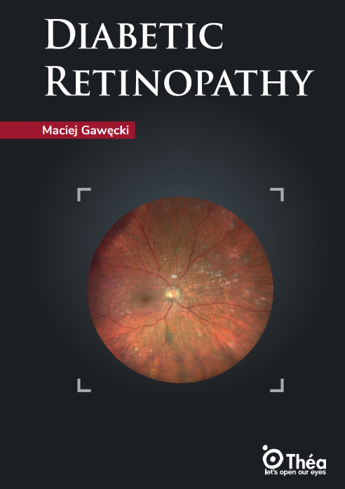
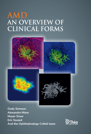
Age-related macular degeneration (AMD) is a leading cause of vision loss in the elderly, affecting the central portion of the retina and progressively impairing central vision. This retinal disease manifests in two primary AMD clinical forms: dry (atrophic) and wet (exudative). Dry AMD, also known as atrophic AMD, involves gradual retinal pigment epithelium degeneration and the accumulation of drusen yellow deposits beneath the retina. This form accounts for approximately 85–90% of AMD cases. It typically progresses slowly and can culminate in geographic atrophy, leading to central vision loss.
Wet AMD, or exudative AMD, constitutes around 10–15% of cases but is responsible for the majority of severe vision impairment. It is characterized by choroidal neovascularization (CNV), where abnormal blood vessels grow under the retina, leaking fluid or blood. Subtypes include type 1 (occult), type 2 (classic), and type 3 (retinal angiomatous proliferation), as well as polypoidal choroidal vasculopathy.
The book also covers intermediate forms such as early AMD, adult-onset vitelliform dystrophy, and reticular pseudodrusen, which are significant due to their potential progression. Each subtype is supported by imaging and case-based descriptions. Prevalence varies with age, genetics, and environmental factors, such as smoking and diet. Understanding the diverse macular degeneration types helps clinicians better anticipate prognosis and customize patient monitoring strategies.
AMD diagnosis relies on multimodal imaging, with optical coherence tomography (OCT) and fluorescein angiography being central to clinical evaluation. OCT provides high-resolution cross-sectional images of retinal layers, identifying drusen, pigment epithelial detachment, and retinal atrophy. Spectral-domain OCT and enhanced depth imaging (EDI-OCT) further help assess choroidal thickness and pachychoroid-related diseases.
Fundus photography color, infrared, and red-free is widely used for documentation and progression tracking. Autofluorescence imaging detects lipofuscin accumulation, indicating pigment epithelium stress or damage. These non-invasive tools are essential for early detection, particularly in asymptomatic intermediate stages.
Fluorescein angiography (FA) remains valuable for visualizing vascular leakage, especially in wet AMD. Indocyanine green angiography (ICGA) complements FA, especially for detecting polypoidal choroidal vasculopathy and occult neovascularization.
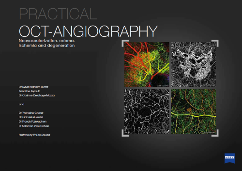
The book also highlights emerging technologies like OCTA (optical coherence tomography angiography), which maps blood flow in retinal and choroidal layers without dye injection. OCTA provides structural and flow-based information, improving detection of quiescent or hidden CNV.
Artificial intelligence and algorithm-based segmentation are increasingly used in image analysis, allowing faster, more accurate interpretation of subtle AMD features.
The primary AMD treatment for wet AMD is anti-VEGF intravitreal injections (e.g., ranibizumab, aflibercept), which inhibit abnormal vessel growth and leakage. These agents significantly reduce the risk of vision loss and are administered regularly based on activity or fixed regimens.
For dry AMD, there is currently no curative treatment, but nutritional supplementation (vitamins C, E, zinc, lutein, omega-3) has shown benefits in slowing progression, as confirmed in AREDS trials. The role of micronutrition is supported by studies emphasizing omega-3 and carotenoid intake.
New therapies in development include complement inhibitors and gene therapies targeting inflammation and neovascular pathways. Anti-inflammatory agents and sustained delivery implants are also under investigation.
Personalized treatment strategies based on genetic profiling (e.g., CFH, ARMS2 polymorphisms) are emerging, especially regarding responsiveness to anti-VEGF therapies. The book also explores how vitelliform dystrophies and other AMD mimickers respond differently, necessitating tailored approaches.
Age-related macular degeneration remains a significant cause of vision impairment, especially in aging populations. Its two major clinical forms dry and wet AMD require distinct diagnostic and therapeutic approaches. Early diagnosis using OCT and angiography is vital to preserve vision and guide timely treatment.
Understanding the full spectrum of AMD clinical forms, including rare presentations and evolving classifications, enhances clinical precision. The book emphasizes the role of emerging technologies like AI imaging and genetics-based predictions in future AMD care. Continued education is essential to keep pace with rapid advancements in imaging, treatment, and research. The Théa Medical Library offers comprehensive ophthalmology book resources, tutorials, and case-based learning, empowering professionals to offer informed and individualized patient care.